| dc.contributor.author | Gönczy, Pierre | - |
| dc.date.accessioned | 2014-01-09T12:03:32Z | |
| dc.date.available | 2014-01-09T12:03:32Z | |
| dc.date.issued | 2003-06 | fr_FR |
| dc.identifier.citation | Gönczy, Pierre ; Mécanismes de division cellulaire : leçons d’un nématode, Med Sci (Paris), 2003, Vol. 19, N° 6-7; p. 735-742 ; DOI : 10.1051/medsci/20031967735 | fr_FR |
| dc.identifier.issn | 1958-5381 | fr_FR |
| dc.identifier.uri | http://hdl.handle.net/10608/4790 | |
| dc.description.abstract | L’embryon du nématode Caenorhabditis elegans représente un système expérimental de choix pour disséquer les mécanismes qui orchestrent les processus de division cellulaire dans un organisme multicellulaire. En effet, un faisceau d’approches cellulaires, génétiques, moléculaires et génomiques permettent d’aborder chez cet organisme nombre de questions fondamentales relatives à la division cellulaire. Certaines des molécules caractérisées chez ce nématode pourraient jouer un rôle important dans d’autres organismes, y compris l’être humain. Dès lors, la recherche fondamentale menée sur l’embryon de C. elegans devrait avoir, à terme, un impact significatif sur le développement de nouveaux outils diagnostiques et thérapeutiques. | fr |
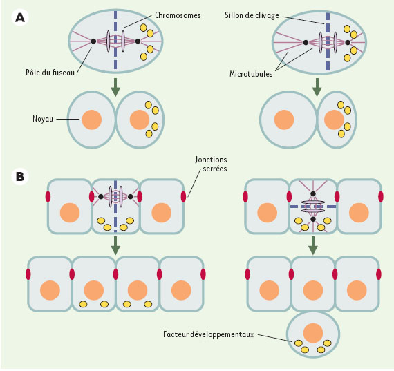
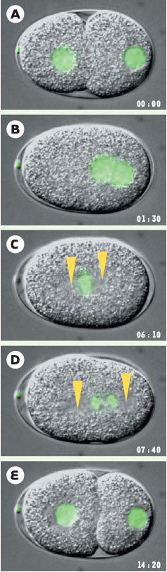
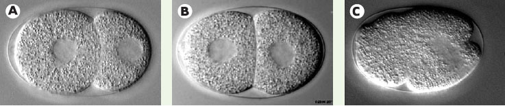
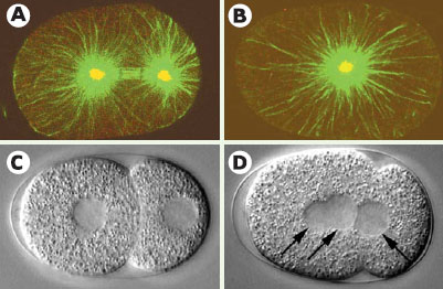
| dc.description.abstract | The mechanisms orchestrating spatial cell division control remain poorly understood. In animal cells, the position of the mitotic spindle dictates cleavage furrow placement, and thus plays a key role in governing spatial relationships between resulting daughter cells. The one-cell stage Caenorhabditis elegans embryo is an attractive model system to investigate the mechanisms underlying spindle positioning in metazoans. In this review, the experimental advantages of this model system for an in vivo dissection of cell division processes are first discussed. Next, three lines of experiments that were conducted to dissect the mechanisms governing spindle positioning in one-cell stage C. elegans embryos are summarized. First, localized laser micro-irradiations were utilized to identify the forces acting on spindle poles during anaphase. This work revealed that there is a precise imbalance of pulling forces acting on the two spindle poles, with the forces acting on the posterior spindle pole being in slight excess, thus explaining the asymmetric spindle position achieved by the end of anaphase. Second, an RNAi-based fonctional genomic screen was carried out to identify novel components required for generating these pulling forces. This uncovered that gpr-1/gpr-2, which encode GoLoco-containing proteins, as well as the previously identified Ga subunits goa- 1/gpa-16, are required for generation of pulling forces on the spindle poles. Third, the zyg-8 locus was identified by mutational analysis to play a distinct role during anaphase spindle positioning. zyg-8 was found to encode a protein related to human Doublecortin, which is affected in patients with neuronal migration disorders. Moreover, ZYG- 8 is a microtubule-associated protein that stabilizes microtubules against depolymerization. Together, these experimental approaches contribute to a better understanding of the mechanisms orchestrating spatial cell division control in metazoan organisms. | en |
| dc.language.iso | fr | fr_FR |
| dc.publisher | EDK | fr_FR |
| dc.relation.ispartof | M/S Revues | fr_FR |
| dc.rights | Article en libre accès | fr |
| dc.rights | Médecine/Sciences - Inserm - SRMS | fr |
| dc.source | M/S. Médecine sciences [ISSN papier : 0767-0974 ; ISSN numérique : 1958-5381], 2003, Vol. 19, N° 6-7; p. 735-742 | fr_FR |
| dc.subject.mesh | Animaux | fr |
| dc.subject.mesh | Caenorhabditis elegans | fr |
| dc.subject.mesh | Protéines de Caenorhabditis elegans | fr |
| dc.subject.mesh | Division cellulaire | fr |
| dc.subject.mesh | Embryon de non mammifère | fr |
| dc.subject.mesh | Développement embryonnaire et foetal | fr |
| dc.subject.mesh | Régulation de l'expression des gènes au cours du développement | fr |
| dc.subject.mesh | Microtubules | fr |
| dc.subject.mesh | Appareil mitotique | fr |
| dc.subject.mesh | Modèles animaux | fr |
| dc.title | Mécanismes de division cellulaire : leçons d’un nématode | fr |
| dc.type | Article | fr_FR |
| dc.contributor.affiliation | ISREC (Institut Suisse de Recherche Expérimentale sur le Cancer), 155, chemin des Bouveresses, CH-1066 Epalinges/Lausanne, Suisse | fr_FR |
| dc.identifier.doi | 10.1051/medsci/20031967735 | fr_FR |
| dc.identifier.pmid | 12942445 | fr_FR |