| dc.contributor.author | Prost-Squarcioni, Catherine | - |
| dc.date.accessioned | 2014-07-03T06:49:33Z | |
| dc.date.available | 2014-07-03T06:49:33Z | |
| dc.date.issued | 2006 | fr_FR |
| dc.identifier.citation | Prost-Squarcioni, Catherine ; Histologie de la peau et des follicules pileux, Med Sci (Paris), 2006, Vol. 22, N° 2; p. 131-137 ; DOI : 10.1051/medsci/2006222131 | fr_FR |
| dc.identifier.issn | 1958-5381 | fr_FR |
| dc.identifier.uri | http://hdl.handle.net/10608/5699 | |
| dc.description.abstract | La peau est complexe, morphologiquement et biochimiquement. Le défi de ces dernières années a été de localiser précisément ses molécules de structure. Dans cet article, l’histologie normale de la peau et des follicules pilo-sébacés est brièvement décrite avec une iconographie en microscopies optique et électronique, afin de mieux comprendre où sont situées les molécules jouant un rôle clé dans la cohésion et la différenciation de l’épiderme, la mélanogenèse, la présentation des antigènes aux lymphocytes T, l’adhérence dermo-épidermique, la résistance et l’élasticité du derme et de l’hypoderme et, enfin, le renouvellement des follicules pileux. Les données présentées ont été établies par la confrontation d’études en peau normale, sur des souris invalidées et chez des patients souffrant de pathologies auto-immunes ou de génodermatoses. L’histologie moléculaire de la peau éclaire sous un autre jour la physiologie de la peau ; elle doit être connue pour valider les essais de thérapie génique ou cellulaire. | fr |
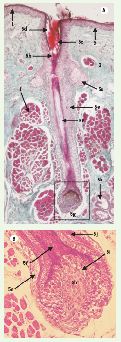
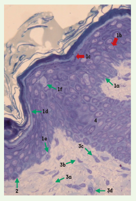
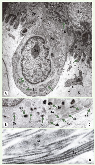
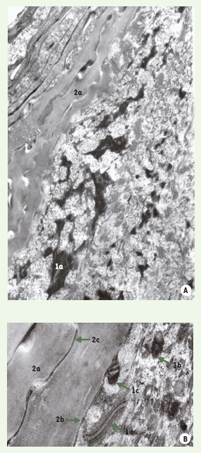
| dc.description.abstract | The skin consists of an outer epidermis, the dermis, and the hypodermis. It includes nerves, blood vessels, glands and hair follicles. Epidermis is a continually renewing, stratified squamous epithelium. It is populated by keratinocytes (80 %) and dendritic cells (20 %) : melanocytes, Langerhans and Merkel cells. In standard histology, keratinocytes are arranged in layers that represent different stages of their differentiation while melanocytes and Langerhans cells appear as clear cells respectively between the basal and the supra-basal cells of epidermis. The Merkel cells cannot be clearly identified. Dendritic processes of the dendritic cells can only be recognized by immunocytochemistry. At the dermal-epidermal junction, a PAS reactive basement membrane follows the contour of the basal cells. Dermis consists of collagenous and elastic fibers embedded into an amorphous ground substance. Fibroblasts, macrophages, mast cells and lymphocytes are its resident cells. Hypodermis is composed of adipocyte lobules defined by fibrous connective tissue septa. Hair follicle consists of 3 parts : the lower portion, from the base of the follicle including hair bulb to the insertion of the arrector pili muscle or buldge ; the isthmus, from the insertion of the arrector pili to the entrance of the sebaceous duct, and the infundibulum, from the entrance of the sebaceous duct to the follicular orifice. The lower portion is composed of the dermal hair papilla, the hair matrix, the hair, and the inner and the outer root sheaths. The hair matrix cells within hair bulb give rise to the hair and to the inner root sheath. With the electron microscope, one can obtain a more detailed view of the characteristic skin structures. Much of them can now be explained in terms of function and in many instances, in correlation with its biochemical composition. An attempt has been made in this paper to precisely give the location of molecules that are relevant in basic skin functions and understanding of auto-immune and genetic diseases. | en |
| dc.language.iso | fr | fr_FR |
| dc.publisher | EDK | fr_FR |
| dc.relation.ispartof | M/S revues | fr_FR |
| dc.rights | Article en libre accès | fr |
| dc.rights | Médecine/Sciences - Inserm - SRMS | fr |
| dc.source | M/S. Médecine sciences [ISSN papier : 0767-0974 ; ISSN numérique : 1958-5381], 2006, Vol. 22, N° 2; p. 131-137 | fr_FR |
| dc.subject.mesh | Adipocytes | fr |
| dc.subject.mesh | Animaux | fr |
| dc.subject.mesh | Tissu conjonctif | fr |
| dc.subject.mesh | Derme | fr |
| dc.subject.mesh | Épiderme | fr |
| dc.subject.mesh | Follicule pileux | fr |
| dc.subject.mesh | Humains | fr |
| dc.subject.mesh | Kératinocytes | fr |
| dc.subject.mesh | Cellules de Langerhans | fr |
| dc.subject.mesh | Mélanocytes | fr |
| dc.subject.mesh | Cellules de Merkel | fr |
| dc.subject.mesh | Glandes sébacées | fr |
| dc.subject.mesh | Peau | fr |
| dc.title | Histologie de la peau et des follicules pileux | fr |
| dc.type | Article | fr_FR |
| dc.contributor.affiliation | Laboratoire d’histologie (EA3410), UFR Léonard de Vinci, 74, rue Marcel Cachin, 93000 Bobigny, France | fr_FR |
| dc.contributor.affiliation | Service de Dermatologie I, Hôpital Saint-Louis, 1, avenue Claude Vellefaux, 75010 Paris, France | fr_FR |
| dc.identifier.doi | 10.1051/medsci/2006222131 | fr_FR |
| dc.identifier.pmid | 16457751 | fr_FR |