| dc.contributor.author | Dufour, Pascal | - |
| dc.contributor.author | Dufour, Suzie | - |
| dc.contributor.author | Castonguay, Annie | - |
| dc.contributor.author | McCarthy, Nathalie | - |
| dc.contributor.author | De Koninck, Yves | - |
| dc.date.accessioned | 2014-07-03T06:52:28Z | |
| dc.date.available | 2014-07-03T06:52:28Z | |
| dc.date.issued | 2006 | fr_FR |
| dc.identifier.citation | Dufour, Pascal ; Dufour, Suzie ; Castonguay, Annie ; McCarthy, Nathalie ; De Koninck, Yves ; Microscopie à deux photons pour l’imagerie cellulaire fonctionnelle : avantages et enjeux ou Un photon c’est bien… mais deux c’est mieux !, Med Sci (Paris), 2006, Vol. 22, N° 10; p. 837-844 ; DOI : 10.1051/medsci/20062210837 | fr_FR |
| dc.identifier.issn | 1958-5381 | fr_FR |
| dc.identifier.uri | http://hdl.handle.net/10608/5876 | |
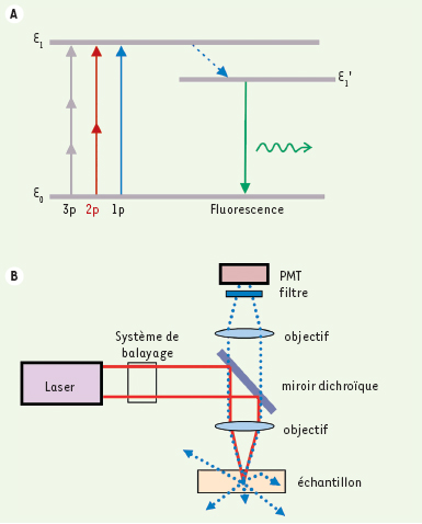
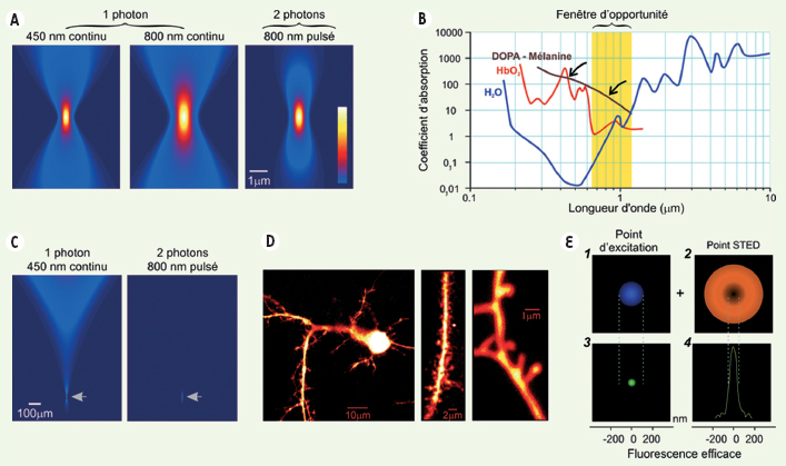
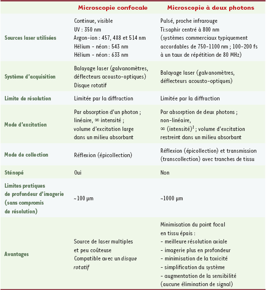
| dc.description.abstract | L’observation de la dynamique des événements moléculaires dans la cellule in situ présente une série de défis, notamment la capacité de suivre ces événements avec le maximum de résolution spatiale et temporelle tout en minimisant l’interférence avec la biologie du tissu et de la cellule. L’exploitation récente d’approches fondées sur l’optique non-linéaire, telle que la microscopie par balayage laser de fluorescence produite par excitation à deux photons, a permis de faire des progrès énormes dans ce domaine, notamment parce qu’elle permet de faire des mesures dans un espace très confiné à l’intérieur du tissu intact et à des profondeurs inaccessibles avec la microscopie linéaire conventionnelle. En minimisant l’excitation indésirable du tissu en dehors du point focal, on améliore la résolution et la sensibilité, on simplifie le système optique et on minimise la phototoxicité. Ces avantages sont à la source du succès de la microscopie à deux photons pour l’imagerie cellulaire fonctionnelle. Des percées récentes en optique/photonique permettent d’envisager d’améliorer davantage la résolution spatiale et temporelle de ce type d’imagerie et la capacité de sonder encore plus profondément dans le tissu pour repousser les limites de la biochimie fonctionnelle et de la biologie cellulaire actuelles. | fr |
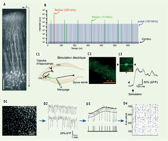
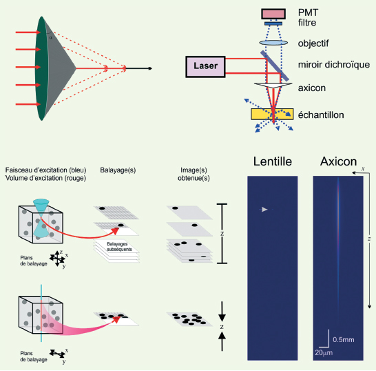
| dc.description.abstract | One of the main challenges of modern biochemistry and cell biology is to be able to observe molecular dynamics in their functional context, i.e. in live cells in situ. Thus, being able to track ongoing molecular events with maximal spatial and temporal resolution (within subcellular compartments), while minimizing interference with tissue biology, is key to future developments for in situ imaging. The recent use of non-linear optics approaches in tissue microscopy, made possible in large part by the availability of femtosecond pulse lasers, has allowed major advances on this front that would not have been possible with conventional linear microscopy techniques. Of these approaches, the one that has generated most advances to date is two-photon laser scanning fluorescence microscopy. While this approach does not really provide improved resolution over linear microscopy in non absorbing media, it allows us to exploit a window of low absorbance in live tissue in the near infrared range. The end result is much improved tissue penetration, minimizing unwanted excitation outside the focal area, which yields an effective improvement in resolution and sensitivity. The optical system is also simplified and, more importantly, phototoxicity is reduced. These advantages are at the source of the success of two-photon microscopy for functional cellular imaging in situ. Yet, we still face further challenges, reaching the limits of resolution that conventional optics can offer. Here we review some recent advances in optics/photonics approaches that hold promises to improve our ability to probe the tissue in finer areas, at faster speed, and deeper into the tissue. These include super-resolution techniques, introduction of non paraxial optics in microscopy and use of amplified femtosecond lasers, yielding enhanced spatial and temporal resolution as well as tissue penetration. | en |
| dc.language.iso | fr | fr_FR |
| dc.publisher | EDK | fr_FR |
| dc.relation.ispartof | M/S revues | fr_FR |
| dc.rights | Article en libre accès | fr |
| dc.rights | Médecine/Sciences - Inserm - SRMS | fr |
| dc.source | M/S. Médecine sciences [ISSN papier : 0767-0974 ; ISSN numérique : 1958-5381], 2006, Vol. 22, N° 10; p. 837-844 | fr_FR |
| dc.subject.mesh | Cellules | fr |
| dc.subject.mesh | Microscopie confocale | fr |
| dc.subject.mesh | Microscopie de fluorescence multiphotonique | fr |
| dc.title | Microscopie à deux photons pour l’imagerie cellulaire fonctionnelle : avantages et enjeux ou Un photon c’est bien… mais deux c’est mieux ! | fr |
| dc.type | Article | fr_FR |
| dc.contributor.affiliation | Centre d’optique, photonique et laser, Département de physique, de génie physique et d’optique, Université Laval, Québec, G1K 7P4 Canada | fr_FR |
| dc.contributor.affiliation | Département de Psychiatrie, Université Laval, Unité de Neurobiologie Cellulaire, Centre de recherché Université Laval Robert-Giffard, 2601, chemin de la Canardière, Québec, G1J 2G3 Canada | fr_FR |
| dc.identifier.doi | 10.1051/medsci/20062210837 | fr_FR |
| dc.identifier.pmid | 17026937 | fr_FR |