| dc.contributor.author | Rémy, P | fr_FR |
| dc.contributor.author | Hantraye, P | fr_FR |
| dc.contributor.author | Samson, Y | fr_FR |
| dc.date.accessioned | 2012-08-23T13:56:33Z | |
| dc.date.available | 2012-08-23T13:56:33Z | |
| dc.date.issued | 1999 | fr_FR |
| dc.identifier.citation | Rémy, P - Hantraye, P - Samson, Y, La tomographie par émission de positons, un outil de recherche fondamentale devenu indispensable à la recherche clinique : l'exemple des greffes neuronales dans la maladie de Parkinson., Med Sci (Paris), 1999, Vol. 15, N° 4; p.490-5 | fr_FR |
| dc.identifier.issn | 1958-5381 | fr_FR |
| dc.identifier.uri | http://hdl.handle.net/10608/1374 | |
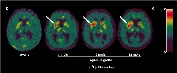
| dc.description.abstract | La neuro-imagerie fonctionnelle, et plus particulierement la tomographie par emission de positons (TEP), est longtemps restee un outil de recherche fondamentale, peu accessible a la recherche therapeutique, notamment en raison de son cout. Neanmoins, dans la maladie de Parkinson, la TEP peut demontrer la survie et le caractere fonctionnel de cellules dopaminergiques foetales implantees dans le striatum de malades parkinsoniens. En effet, en mesurant la fonction dopaminergique avec la L-Dopa marquee au fluor 18 radioactif, la TEP a permis de montrer l' augmentation du captage de ce traceur chez les patients apres la greffe, parallelement a une amelioration clinique lorsque celle-ci s' est manifestee. Seul outil capable de mesurer directement in vivo l' evolution de la survie des neurones dopaminergiques dans la maladie de Parkinson, la TEP a, depuis, acquis un role important dans les essais a grande echelle de traitements neuroprotecteurs. | fr |
| dc.description.abstract | Functional neuroimagery and more particularly positron emission tomography (PET) has long been restricted to fundamental neuroscientific research, with little applications in clinical research mainly because of its high cost. However, in parkinsonian patients treated using fetal neuronal grafts, PET has demonstrated the survival and function of dopaminergic cells implanted into the striatum. By measuring the dopaminergic function using L Dopa labeled with radioactive fluorine 18, PET has shown an increased uptake of the tracer after transplantation, which parallels the clinical improvement of patients. Since only PET is able to monitor in vivo the degeneration of dopaminergic cells of parkinsonian patients, it a has become a tool in evaluating neuroprotective agents. PET is also used for the evaluation of new treatments in other neurodegenerative diseases such as, for example, Huntington's disease. | en |
| dc.language.iso | fr | fr_FR |
| dc.publisher | Masson, Paris | fr_FR |
| dc.rights | Article en libre accès | fr |
| dc.rights | Médecine/Sciences - Inserm - SRMS | fr |
| dc.source | M/S. Médecine sciences [revue papier, ISSN : 0767-0974], 1999, Vol. 15, N° 4; p.490-5 | fr_FR |
| dc.title | La tomographie par émission de positons, un outil de recherche fondamentale devenu indispensable à la recherche clinique : l'exemple des greffes neuronales dans la maladie de Parkinson. | fr |
| dc.title.alternative | Positron emission tomography, a new clinical research tool : the example of neuronal grafts in Parkinson's disease | fr_FR |
| dc.type | Article | fr_FR |
| dc.contributor.affiliation | CEA, Service hospitalier Frederic-Joliot, Departement de recherche medicale, 91406 Orsay, France; Departement de neurosciences medicales, CHU Henri-Mondor, 51, avenue du Marechal-de-Lattre-de-Tassigny, 94010 Creteil, France; Ura CEA Cnrs 2210, Service hospitalier Frederic-Joliot, Departement de recherche medicale, 91401 Orsay, France | - |
| dc.identifier.doi | 10.4267/10608/1374 | |