


Représentation cérébrale de l'expérience subjective de la douleur chez l'homme.
Résumé
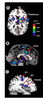
Une stimulation nociceptive mobilise une multitude de
structures cérébrales. Cette activation cérébrale est à l’origine
de l’expérience subjective de la douleur et reflète vraisemblablement
ses dimensions sensorielle et affective. Chez
l’homme, l’application de stimulations douloureuses produit
une augmentation multifocale et stéréotypée du débit sanguin
cérébral régional, détectée dans des études d’imagerie
cérébrale tomographique par émission de positons. L’activation
corticale est observée principalement dans l’hémisphère
controlatéral au site de stimulation, dans les aires somato-sensorielles
primaire et secondaire, dans l’insula et dans le
cortex cingulaire antérieur. Des foyers d’activation sous-corticale
sont également observés. La réponse des régions corticales
est directement proportionnelle à l’expérience subjective
de douleur et elle est modifiée par des interventions
cognitives altérant la perception de l’intensité ou le désagrément
lié à la douleur, par exemple lors de suggestions
hypnotiques ou de fixations différentielles de l’attention. A threat to the integrity of the organism by nociceptive stimulation activates multiple cerebral structures. The cerebral activation gives rise to a subjective experience of pain that comprises a sensory dimension (e.g. intensity) and an affective dimension (e.g. unpleasantness). Brain imaging studies using positron emission tomography (PET) in humans demonstrate a multifocal and stereotyped pattern of activation in response to painful stimulation applied to the skin, muscle or viscera. The cortical activation is observed mainly in the contralateral hemisphere, in primary and secondary somatosensory areas (S1 and S2), in the Insula of Reil, and in the anterior cingulate cortex (ACC). Sub-cortical foci of activation are found in brain stem, thalamus, hypothalamus, lenticular nuclei, and cerebellum. The activity measured in cortical areas is proportional to the subjective experience of pain, and the modulation of the sensory or affective dimensions of pain by cognitive interventions produces characteristic changes in cortical activity. Activity in ACC is larger following hypnotic suggestions to increase pain affect than following suggestions to decrease pain affect. Similarly, activity in S1 is larger following hypnotic suggestions to increase pain intensity than following suggestions to decrease pain intensity. Moreover, cerebral activity particularly in S1 and the perception of pain intensity, increase in parallel when attention is directed to the painful stimuli during a task of intensity discrimination, and decrease when attention is directed toward concurrent auditory stimuladon. Pain is subserved by a vast network of cortical structures, each of which participates differently in the multiple aspects of the experience. Results of our studies suggest at least a partial functional segregation of regions involved in the sensory and affective dimensions of pain. A threat to the integrity of the organism by nociceptive stimulation activates multiple cerebral structures. The cerebral activation gives rise to a subjective experience of pain that comprises a sensory dimension (e.g. intensity) and an affective dimension (e.g. unpleasantness). Brain imaging studies using positron emission tomography (PET) in humans demonstrate a multifocal and stereotyped pattern of activation in response to painful stimulation applied to the skin, muscle or viscera. The cortical activation is observed mainly in the contralateral hemisphere, in primary and secondary somatosensory areas (S1 and S2), in the Insula of Reil, and in the anterior cingulate cortex (ACC). Sub-cortical foci of activation are found in brain stem, thalamus, hypothalamus, lenticular nuclei, and cerebellum. The activity measured in cortical areas is proportional to the subjective experience of pain, and the modulation of the sensory or affective dimensions of pain by cognitive interventions produces characteristic changes in cortical activity. Activity in ACC is larger following hypnotic suggestions to increase pain affect than following suggestions to decrease pain affect. Similarly, activity in S1 is larger following hypnotic suggestions to increase pain intensity than following suggestions to decrease pain intensity. Moreover, cerebral activity particularly in S1 and the perception of pain intensity, increase in parallel when attention is directed to the painful stimuli during a task of intensity discrimination, and decrease when attention is directed toward concurrent auditory stimuladon. Pain is subserved by a vast network of cortical structures, each of which participates differently in the multiple aspects of the experience. Results of our studies suggest at least a partial functional segregation of regions involved in the sensory and affective dimensions of pain.
Pour citer ce document
Rainville, P ; Duncan, GH ; Bushnell, MC, Représentation cérébrale de l'expérience subjective de la douleur chez l'homme., Med Sci (Paris), 2000, Vol. 16, N° 4; p.519-27