


L'induction neurale chez les vertébrés : le cerveau par défaut.
Résumé
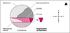
Classiquement, le système nerveux de l’embryon d’amphibien
nait, au cours de la gastrulation, d’interactions inductrices
entre le mésoderme dorsal et l’ectoderme qui le
recouvre ; elles mettent en jeu trois facteurs diffusibles, Noggin,
Follistatin et Chordin, sécrétés par l’organisateur de Spemann.
Les études actuelles montrent que l’induction neurale
semble, en outre, liée à la levée d’une inhibition des cellules
à devenir neurales : c’est le modèle dit du cerveau par
défaut. On a montré que la protéine BMP4 (bone morphogenetic
protein), un autre membre de la superfamille TGFβ,
induit la différenciation épidermique et inhibe la spécification
neurale ; à l’inverse, toute interférence avec la voie de
transmission du signal BPM4, en particulier par Noggin, Follistatin
et Chordin, provoque l’induction neurale. Ces mécanismes
moléculaires sont conservés au cours de l’évolution et
leur étude pourrait conduire à des développements thérapeutiques
intéressants dans les maladies neurodégénératives. Classical experiments performed in the amphibian embryo established that the vertebrate nervous system arises, during gastrulation, from inductive interactions between the dorsal mesoderm and the overlaying ectoderm. Transplantation of the gastrula dorsal lip in the ventral side of a host embryo results in a twin embryo with a complete ectopic dorsal axis in the ventral side. Lineage tracing experiments have shown that while the axial mesoderm in the ectopic axis is derived from the explant itself, the nervous system is derived from the ventral ectoderm of the host embryo which otherwise would have formed epidermis. While these experiments demonstrated that gastrula dorsal mesoderm, termed the 'organizer', can change the fate of ectodermal cells from epidermal to neural, only recently has the molecular nature of these organizer signals begun to be elucidated. Experiments performed in amphibians have led to the characterization of three diffusible factors, noggin, follistatin and chordin, all localized in the organizer, the embryonic source of neural inducing signals. These factors can change the fate of ectodermal cells from epidermal to neural directly without concomitant mesoderm induction. Based on studies with TGF-beta antagonists, such as the truncated activin receptor (Delta1XAR1) and follistatin, we proposed that the formation of the nervous system in vertebrates is under negative control and requires the inhibition of an inhibitor, activin or a related factor, which can be antagonized with Delta1XAR1 and follistatin. We called this the 'default model' of vertebrate neural specification. This model provides the molecular link between the observation made by classical experimental embryologits and more recent experiments involving dissociated animal caps. Several groups have shown that animal caps, which give rise to epidermis if kept intact, will form neural tissue if the cells of the explants are dissociated for several hours. These results suggest that dilution of a diffusible 'neural inhibitor' from the explants provides another way to eliminate a neural inhibitor. Later experiments demonstrated that BMP4, another member of TGF-beta superfamily, can inhibit neural specification and induce epidermis in dissociated Xenopus embryonic ectodermal cells, pointing to the involvement of BMP4 in cell fate decisions in the context of the ectoderm. Recent experiments with chordin, noggin and dominant negative forms of BMP4 ligands and receptors support this hypothesis, thus providing additional evidence for the default model and for neuralization being under inhibitory control. Interference with TGF-beta signaling in a mammalian embryonic cell line also leads to the specification of neural cells, and BMP4 inhibits the retinoic acid induced neural differentiation suggesting that the molecular strategy behind neural determination has been conserved throughout vertebrate evolution. Thus, the model emerging from these results proposes that the default state of Xenopus ectoderm is neural, that BMP4, and possibly other members of the TGF-beta family inhibit neuralization, and that BMP4 induces cells to become epidermal. The isolation and characterization of proteins involved in neural and epidermal induction has immediate health related consequences. Factors which induce neural differentiation in ectodermal and mesodermal cells may provide insights into regeneration of neural tissue in adults and, in the long term, may provide therapy for diseases involving loss of neuronal cells, including stroke and neurodegenerative diseases. In addition, the characterization of an epidermal inducer may lead to treatments for skin injuries and diseases. The collection of this type of knowledge will provide the basis for development of rational treatments. [References: 45]
Pour citer ce document
Honoré, E ; Hemmati-Brivanlou, A, L'induction neurale chez les vertébrés : le cerveau par défaut., Med Sci (Paris), 1997, Vol. 13, N° 2; p.192-200